Menlo Osteopathy
MUSCULOSKELETAL MEDICINE
A Whole - Person Pursuit

Physical Medicine and Rehabilitation

Physical Medicine and Rehabilitation
The end goal is the same as it is in Osteopathy “to restore function.” However, the approach by which this is accomplished may be different. Perhaps more direct and sorted. Given my training, I tend to marry the two approaches and enhance one by the other. I view as you, as an individual have an option, the opportunity to choose how to proceed with restoring your dis-ease. Physical Medicine and Rehabilitation specialists’ (“physiatrists”) primary goal is to rebuild function in a wide range of conditions. Those include sports, motor vehicle injuries, neurologic disorders and other physical disabilities. The common thread among all is impaired function. Physiatrists’ approach to treat musculoskeletal impairments and increase function is usually multifaceted through detail physical exams, physical therapies, medications, assistive devices and non-surgical interventions.


.jpg)
Interventional Approach to Injury, Pain and Dysfunction
Ultrasound (US) guidance
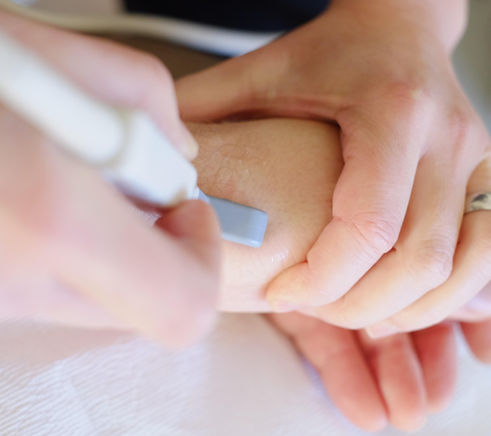
Interventional pain procedures have been conducted with and without visualization for decades. Fluoroscopy proved to be the most common imaging tool up to recent years, when ultrasound (US) has emerged. Visualization is key when considering diagnostic accuracy and targeted treatment, however interventional pain procedures have been successfully conducted utilizing surface landmarks alone. Nothing wrong with that! However, pain medicine guidelines suggest that most procedures require image guidance. Ultrasound allows for identification of pathology, visualization of the needle in real time, accuracy of needle placement and dynamic assessment of musculoskeletal pain. Above all, it eliminates radiation exposure. In short, US guidance enhances both; diagnostic and therapeutic purposes with regards to structural abnormalities. Dr. Jarosz uses an ultrasound to evaluate musculoskeletal pathology which if untreated may lead to chronic pain. She conducts interventional pain procedures in addition to osteopathic manipulative treatment (OMT).

Interventional Pain Procedures
Intra-Articular Join Injections
A combination of an anesthetic agent such as lidocaine and a corticosteroid is injected into a joint such as shoulder, knee, finger, wrist, ankle, toe. The purpose would be to manage long standing pain and/or to improve exercise or physical therapy tolerance.
Classic Corticosteroid injections
Neuromusculoskeletal pathology have traditionally responded well to corticosteroid injections. These agents decrease inflammation, reduce swelling, thus reduce pain. Corticosteroids are useful in numerous disorders spanning from general osteoarthritis to elevated intra-regional pressure conditions such as peripheral nerve neuropathies (carpal tunnel syndrome), compressed spinal nerves and tendon pathologies, including shoulder impingement. Dr. Jarosz performs ultrasound-guided injections for a multitude of musculoskeletal conditions, including shoulder, hip dysfunctions, calcific tendonitis, tendon contractures, nerve entrapments and joint pathology.


Hyaluronic Acid (HA) Injections
Hyaluronic Acid is a molecule found naturally throughout the human body; in our joints, connective tissue and skin. Depending on where
it is in the body, it has very distinct functions. In the skin, it hydrates.
In the connective tissue, it hydrates and cushions the living cells.
In joints, it acts as a lubricant and shock absorbent, thanks to its
ability to bind and retain water molecules. This in turn leads to more space within the joint, less grinding and less pain. It is an important component of joint fluid (also known as synovial fluid), which provides lubrication and
cushioning for normal, healthy knees. HA is especially beneficial for mild to moderate knee osteoarthritis where the production and
quality of hyaluronic acid is inferior in comparison to non-arthritic
knee joints, and indicated for individuals who failed conservative treatment such as physical therapy and/or oral analgesics.
Hyaluronic Acid is safe and well tolerated as intra-articular knee joint injections. Its formulations and dosing vary, ranging from a single to a multiple-injection regimens. HA should cause no interactions with other therapeutic injections, immunizations or
your usual oral medications.

In the case of knee osteoarthritis the endogenous hyaluronic acid breaks down and reduces in concentration. This process weakens its natural elastic properties, which can propagate cartilage degradation and increase joint pain. I partner with brands which strive to mimic the synthetic HA composition to the naturally synthesized hyaluronic acid in your body, but yet boost its natural concentration and chemical stability.

Prolotherapy
Prolotherapy is an umbrella term for different types of pro-inflammatory injectants. Those include Traditional Prolotherapy, an active ingredient dextrose (a form of sugar) which triggers a local regenerative response. Others include Platelet Rich Plasma (PRP) and Stem-Cell Injections. Prolotherapy induce inflammation at the site of needed healing and activates formation of new connective tissue. This is a similar process to healing a skin cut. It alleviates chronic pain, joint laxity, and many other musculoskeletal issues without the risks or long recovery time of surgery. There is no necessary recovery time and regular activity can be maintained between treatments.


Extracorporeal Shock-Wave Therapy (ESWT)
Extracorporeal shock waves are sound waves generated outside of the body, originally used for fragmentation of kidney stones. ESWT has a wide variety of applications in modern medicine, including orthopedic/musculoskeletal pathology. Shock waves are sound (acoustic) waves and capable of transmitting energy from point of generation to remote regions (from point A to point B). They require a medium such as water or air for propagation. Human tissues consist primarily of water with very similar transmission properties and shock waves are transmitted to the biological tissue without substantial loss. Most shock wave therapy result in increased blood flow and enhanced metabolic activity, leading to healing.
Mechanical energy in the form of an acoustic pressure wave is transmitted to the body tissue, such as a painful region by means of transmitters. These pressure waves are very helpful in treatment of musculoskeletal disorders. Radial pressure waves generate oscillations in tissue which lead to improved microcirculation and increased metabolic activity. ESWT induces new collagen and vascular formation, thus improves symptoms and function in clinical cases such as Dupuytren's Contracture, mild-to-moderate carpal tunnel syndrome, diabetic foot ulcer (DFU), chronic plantar fasciitis, tendinosis and first carpometacarpal (CMC) joint osteoarthritis. This in turn improves acute or chronic pain and improves overall function.

Exercise
Exercise is Medicine! There are various exercise prescriptions. However, a blanketed statement of any movement is better than none and numerous benefits arise from exercise. These range from building muscle, bone, balance, preventing falls, injury, dis-ease, to enhancing soft tissue collagen production. Most sports medicine and musculoskeletal conditions may be managed non-surgically. The foundation for this form of treatment is to improve the quality of movement through physical therapy. The goals for each patient should be taken into account to provide patient-centered care.
Physical therapy is the first-line in the treatment and management of injuries. Initial goals may include reducing irritation to tissue, joints and muscles followed by restoring range of motion, strength and progression to restoring the full kinetic chain (coordination of movements in adjacent segments such as spine, hip, knee and foot/ankle of the lower extremity).
